A — Asymmetry
One half of a mole or pigmented lesion looks different from the other.
Private Skin Cancer Assessment — Newmarket
In-person clinical assessment and dermoscopy for a mole, patch, sore or growth that concerns you — with biopsy, histology, selected skin-cancer surgery and coordinated onward referral when needed.
CQC-registered clinic · Clinician-led assessment · No GP referral required for self-pay care
CQC Registered
Rated "Good" at inspection
5.0 on Google
137 patient reviews
Medical practitioners only
Doctors, nurse prescribers & dentists
Honest consultations
No obligation to proceed
What to look for — 01
Many moles, marks and growths are harmless. What matters is noticing a lesion that is new, changing, unusual for you or behaving differently from the rest. The ABCDE guide can help you recognise changes in pigmented lesions, but not every melanoma follows every letter and not all skin cancers are moles.
Basal cell and squamous cell carcinomas may appear as a persistent growth, scaly or crusted patch, a sore that bleeds or does not heal, or an area that repeatedly heals and returns. These changes deserve clinical assessment rather than online or photographic self-diagnosis.
Seek prompt advice: contact your GP, NHS 111 where appropriate, or a qualified skin-cancer service if a lesion is changing, bleeding, crusting, painful, itchy or not healing. An urgent referral does not mean that you definitely have cancer.
One half of a mole or pigmented lesion looks different from the other.
Edges are irregular, blurred, notched or changing rather than smooth and even.
There is a mix of colours or a new change in colour, including black, brown, red, pink, blue or white areas.
The lesion is enlarging or is around 6 mm or more, although skin cancers can be smaller.
A mole or lesion is changing in size, shape, colour, surface, sensation or behaviour over time.
Your consultation — 02
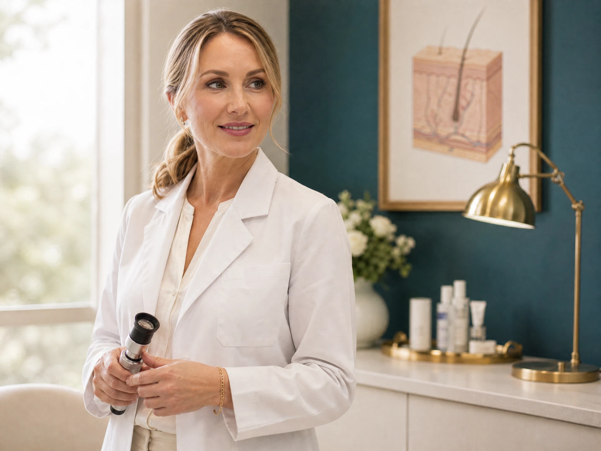
Unlike an image-only reporting service, your appointment is an in-person clinical consultation. We consider the lesion, its history, your symptoms and relevant risk factors, then use dermoscopy where appropriate. You leave with an explanation and a documented next step rather than an isolated photograph or score.
Clinical assessment and dermoscopy of the lesion or lesions causing concern.
Biopsy or excision with histology when tissue diagnosis is required.
Selected BCCs and some SCCs removed under local anaesthetic when appropriate.
Clear onward referral for suspected melanoma, complex sites or specialist treatment.
Dermoscopy & histology — 03
A dermatoscope uses magnification and specialised light to reveal colours and structures beneath the skin surface. NICE recommends dermoscopy during specialist assessment of suspected melanoma because it can improve distinction between benign and malignant lesions when used by a suitably trained clinician.
Dermoscopy informs the decision; histology confirms the tissue diagnosis. If a biopsy or excision is appropriate, the specimen is examined by a pathologist. The report identifies the lesion and, where relevant, comments on margins. We explain the result and whether any further treatment or monitoring is needed.
Treatment at our clinic — 04
We do not limit minor surgery to benign lesions. Selected basal cell carcinomas and some squamous cell carcinomas can be removed at our CQC-registered clinic under local anaesthetic, followed by histology, wound review and results discussion. Suitability depends on diagnosis, size, location, recurrence risk, patient health and the type of surgery required.
Minor surgery, methods and feesOnward hospital care — 05
Suspected melanoma, high-risk or recurrent disease, complex anatomical sites, and lesions that may need Mohs surgery, plastic surgery or multidisciplinary management are referred onward. We can provide the receiving local NHS or private hospital service with the consultation findings, dermoscopic information and histology available, helping the next team understand what has already been assessed.
Dermatology careFees & insurance — 06
A skin cancer check with our GP with Special Interest in Dermatology is £180; an appointment with a Dermatology Clinical Nurse Specialist is £150. If specialist hospital assessment is needed, we advise on referral. Biopsy and surgery fees depend on the method, lesion and complexity; you receive a quotation before treatment. Histology and planned follow-up are included with the excision prices shown on our Minor Surgery page.
Aviva, WPA, TRICARE Overseas and other private plans may reimburse eligible medically necessary care, subject to referral, pre-authorisation and policy terms. Confirm cover before booking. Cosmetic lesion removal is self-funded. We can provide itemised invoices and supporting clinical documentation.
Read our insurance guideConcerned about a mole or lesion?
No GP referral is needed for self-pay care. If using insurance, obtain any required referral and authorisation first.
FAQ
Can't find your answer? Call us on 01638 476423 — our team will be glad to help.
Clinical information sources: NICE guidance on dermoscopy and suspected cancer referral; NHS guidance on melanoma and non-melanoma skin-cancer symptoms and treatment; Royal Marsden guidance on skin-cancer diagnosis; Cancer Research UK patient information. Page last reviewed 5 September 2026.
Learn more in the Education Station